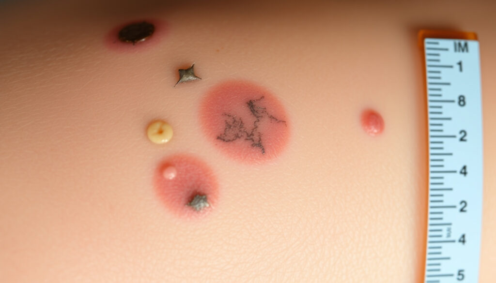
Pictures of Cancerous Moles – Early Signs Identification Guide
Recognising the early warning signs of skin cancer can be a matter of significant clinical importance. Whether you are checking a suspicious mole after a summer of sun exposure or monitoring an existing spot for changes, understanding what potentially cancerous growths look like empowers you to seek timely professional evaluation. This guide brings together authoritative descriptions and visual reference points from leading dermatological sources to help you distinguish between harmless moles and those that warrant medical attention.
Skin cancer remains one of the most prevalent forms of cancer globally, yet when identified at an early stage, treatment outcomes are frequently highly favourable. The three primary types—basal cell carcinoma, squamous cell carcinoma, and melanoma—each present distinctive visual characteristics that distinguish them from ordinary skin markings. Learning to recognise these patterns does not replace professional diagnosis but can serve as a valuable first step toward appropriate care.
What Do Early Stage Pictures of Cancerous Moles Look Like?
Early stage skin cancer frequently manifests as subtle alterations to existing moles or the emergence of new skin markings that warrant closer examination. According to the Medical News Today overview of early-stage skin cancer, common presentations include irregular spots, sores that fail to heal within several weeks, scaly patches, and moles that undergo measurable changes in size, colour, or texture.
The most widely recognised framework for evaluating suspicious moles involves five criteria: Asymmetry, Border irregularity, Colour variation, Diameter exceeding 6mm, and Evolution over time. These features, when present individually or in combination, indicate that a mole may warrant dermatological assessment.
Four-Item Overview: Core Warning Signs
- Early Stage Signs: Asymmetry and irregular, blurred, or notched borders distinguish potentially problematic moles from their benign counterparts.
- Common Locations: Lesions frequently appear on sun-exposed areas including the legs, back, face, neck, and ears.
- Types Covered: Melanoma, basal cell carcinoma, and squamous cell carcinoma each display characteristic early appearance patterns.
- Key Framework: The ABCDE rule provides a systematic approach to self-examination that can prompt timely professional consultation.
Key Insights on Identifying Cancerous Moles
- Melanoma spots may display uneven pigmentation encompassing shades of tan, brown, black, white, or red.
- Basal cell carcinoma often appears as a pearly, translucent, shiny bump that may crust, bleed, or resist healing.
- Squamous cell carcinoma in early stages presents as rough, scaly patches or wart-like growths on sun-damaged skin.
- Changes in size, shape, colour, or surface texture occurring over weeks or months constitute an evolving mole requiring evaluation.
- Spots larger than 6mm—approximately the dimensions of a standard pencil eraser—merit closer inspection.
- Moles exhibiting multiple colours or uneven colour distribution should be examined by a dermatologist.
- The absence of pain or irritation does not exclude malignancy; many early skin cancers remain asymptomatic.
| Visual Feature | Benign Characteristic | Cancerous Indicator |
|---|---|---|
| Symmetry | Both halves mirror each other | One half differs from the other |
| Border | Smooth, well-defined edge | Irregular, blurred, or notched edges |
| Colour | Single uniform shade | Multiple shades or uneven distribution |
| Diameter | Smaller than 6mm | Larger than 6mm |
| Evolution | Remains unchanged over time | Changes in size, shape, or colour |
| Surface | Smooth, flat or slightly raised | Rough, scaly, crusted, or bleeding |
Melanoma Pictures on Legs, Back, and Face
Melanoma represents the most serious form of skin cancer, yet when identified before it penetrates deeper skin layers, cure rates remain remarkably high. The Mayo Clinic resource on melanoma identification documents that early-stage melanomas typically manifest as asymmetric spots exceeding 6mm in diameter, often displaying irregular borders and colour variation that distinguishes them from ordinary moles.
Melanoma on the Legs
The lower extremities rank among the most frequent sites for melanoma development in adults. Photographs from dermatological collections illustrate how leg melanomas often appear as irregular moles with asymmetric shapes and multiple colour gradients, ranging from light brown to dark black. Healthcare professionals recommend regular examination of the entire leg surface, including areas that typically remain concealed by clothing.
Melanoma on the Back
Melanomas developing on the back frequently escape early detection due to the difficulty of self-examination in this location. The Skin Cancer Foundation’s image gallery demonstrates how trunk melanomas may present as irregular spots with uneven pigmentation, sometimes beginning as small lesions that gradually expand and develop additional warning features.
Melanoma on the Face
Facial melanomas account for a substantial proportion of cases and may develop from existing moles or appear as new growths. Images from medical sources reveal that facial melanomas can range from pigmented lesions displaying classic ABCDE features to amelanotic melanomas that appear pink, reddish, or colourless—a variant that may escape notice without careful inspection.
Research from the Skin Cancer Foundation confirms that melanoma can develop on any skin tone. On darker skin, melanomas may appear more pigmented with a pearly or translucent quality, and they frequently occur in less sun-exposed locations such as the palms, soles, and under nails. This underscores the importance of comprehensive skin examinations regardless of complexion.
Types of Skin Cancer Pictures Including Basal Cell Carcinoma
Beyond melanoma, other forms of skin cancer possess distinct visual signatures that aid in recognition. Understanding these variations equips individuals with broader awareness when conducting skin self-examinations.
Basal Cell Carcinoma Visual Characteristics
Basal cell carcinoma stands as the most commonly diagnosed form of skin cancer worldwide. GoodRx clinical descriptions detail how BCC typically presents as a pearly or translucent bump with a smooth, rolled border, often described as resembling a mole or flesh-coloured wart. Growths may appear flesh-coloured, pink, or exhibit pigmentation, and commonly develop on sun-exposed regions including the face, neck, ears, and trunk.
Photographic references from Miiskin’s dermatological image collection illustrate how BCC may ulcerate, bleed intermittently, or form a sore that fails to heal completely over several weeks. While BCC grows slowly and rarely spreads to distant sites, early intervention remains important for optimal cosmetic and functional outcomes.
Squamous Cell Carcinoma Visual Characteristics
Squamous cell carcinoma often develops on skin that has sustained cumulative sun damage over years. The American Academy of Dermatology picture gallery demonstrates how SCC may appear as rough, scaly red or pink patches, dome-shaped wart-like growths, or craters with elevated edges. Lesions frequently arise on the face, ears, lips, backs of hands, and lower legs.
Any spot that bleeds spontaneously, fails to heal within four to six weeks, or rapidly changes in appearance should prompt urgent dermatological assessment. While many non-cancerous conditions mimic these features, only a professional examination with potential biopsy can confirm or exclude malignancy.
Actinic Keratosis: The Pre-Cancerous Stage
Actinic keratosis represents a precancerous condition that may progress to squamous cell carcinoma if left unaddressed. These rough-textured, pinkish patches typically emerge on sun-exposed areas such as the hands, forearms, and face. Medical image collections show how actinic keratoses often feel gritty or sandpaper-like to the touch, providing a tactile clue that supplements visual inspection.
Timeline: How Moles Progress to Cancer
Understanding the progression pathway from normal skin to malignancy provides valuable context for why early detection carries such significance.
- Normal Mole: A uniform, symmetric growth with smooth borders and consistent colouring, typically smaller than 6mm, remaining stable over time.
- Dysplastic Nevus: An atypical mole displaying some concerning features such as irregular borders or mixed pigmentation, yet not meeting criteria for melanoma. These require monitoring but are not yet malignant.
- Early-Stage Melanoma: A mole exhibiting one or more ABCDE warning signs—typically asymmetry, irregular borders, multiple colours, diameter exceeding 6mm, or observable change over time. This stage remains highly treatable.
- Locally Advanced Melanoma: Cancer cells have penetrated deeper skin layers but remain confined to the immediate area. Treatment options remain numerous and outcomes favourable with prompt intervention.
- Metastatic Melanoma: Cancer cells have spread to lymph nodes or distant organs, requiring more intensive treatment approaches. This stage underscores the critical importance of detecting changes before metastasis occurs.
Benign vs Cancerous Moles: Spotting the Difference
Distinguishing between harmless moles and potentially malignant growths requires careful attention to specific characteristics, though definitive diagnosis always necessitates professional medical evaluation.
Benign moles typically maintain uniform colouration, smooth borders, and consistent dimensions over years. Cancerous growths more frequently display asymmetry, multiple pigment shades, irregular edges, diameters exceeding 6mm, or documented change over time. The presence of any single warning feature warrants professional assessment, while combinations of multiple features increase concern significantly.
It bears emphasis that many non-cancerous conditions can mimic the appearance of skin cancer. Irritated or inflamed moles, seborrheic keratoses, and various benign growths may display features that appear concerning upon initial inspection. Conversely, some melanomas—in particular the amelanotic variant—may lack the pigmentation typically associated with malignancy.
Skin Cancer Images from NHS and Official Sources
Several authoritative health organisations maintain image galleries and educational resources that supplement clinical descriptions with visual references. The NHS melanoma skin cancer symptom page provides UK-specific guidance aligned with international detection frameworks, while the Centers for Disease Control and Prevention skin cancer information portal offers structured educational content on prevention and early recognition.
The World Health Organization fact sheet on skin cancers contextualises the global burden of this disease category, reinforcing why public awareness of early warning signs serves as an important public health measure.
The Cancer.org skin cancer image gallery provides extensive photographic references spanning multiple cancer types, stages, and skin tones—valuable for individuals seeking to understand the range of presentations across diverse populations.
What to Do If You Spot a Suspicious Mole
Discovering a potentially concerning skin lesion naturally prompts questions about appropriate next steps. Pinched Nerve in Neck – Symptoms, Causes and Treatments addresses how unrelated conditions may sometimes cause concern that prompts medical evaluation, reinforcing the principle that any new or changing skin feature warrants professional assessment.
The recommended approach involves conducting a systematic self-examination using the ABCDE framework, documenting any findings with photographs for future comparison, and scheduling an appointment with a dermatologist for persistent or concerning spots. During the consultation, the dermatologist may perform a dermoscopy examination using magnified imaging, and if warranted, a biopsy to obtain tissue for laboratory analysis.
Individuals with numerous moles, personal history of skin cancer, or significant sun exposure may benefit from regular dermatologist check-ups independent of self-detected concerns. Signs of Low Iron – Recognize Deficiency Early demonstrates how attention to bodily warning signs—whether skin-related or systemic—supports proactive health management.
Summary
Recognising potentially cancerous moles relies on understanding characteristic warning signs rather than attempting to memorise every possible presentation. The ABCDE framework—asymmetry, border irregularity, colour variation, diameter exceeding 6mm, and evolution—provides a practical tool for self-assessment. Early-stage skin cancer pictures from authoritative sources illustrate how these principles manifest across different cancer types and body locations, from facial basal cell carcinomas to leg melanomas. Professional evaluation remains essential for any spot that raises concern, as timely diagnosis significantly improves treatment outcomes across all skin cancer types.
Frequently Asked Questions
What do cancerous moles look like?
Cancerous moles often display asymmetry, irregular borders, multiple colours, diameter exceeding 6mm, or visible changes over time. Basal cell carcinoma may appear as a pearly bump, squamous cell carcinoma as a scaly patch, and melanoma as an irregular, multi-coloured spot. Any concerning mole warrants professional dermatological assessment.
How can I tell if a melanoma is developing on my leg?
Melanoma on the legs frequently appears as an irregular mole with asymmetric shape and uneven colour distribution ranging from light brown to dark black. Check the entire leg surface regularly, using mirrors or photographs for hard-to-see areas, and monitor for any new or changing spots.
What does basal cell carcinoma look like in early stages?
Early basal cell carcinoma typically presents as a pearly, translucent, shiny bump or nodule, often with a smooth, rolled border. Lesions may be flesh-coloured, pink, or slightly pigmented and commonly develop on sun-exposed areas including the face and neck.
Can skin cancer develop on darker skin tones?
Yes, skin cancer including melanoma can develop on any skin tone. On darker skin, melanomas may appear more pigmented or pearly and frequently occur on less sun-exposed areas such as the palms, soles, and under nails.
When should I see a doctor about a mole?
Schedule a dermatological appointment for any mole displaying asymmetry, irregular borders, multiple colours, diameter exceeding 6mm, or changes over time. Also seek evaluation for spots that bleed, crust, fail to heal within several weeks, or cause any discomfort.
What is the difference between actinic keratosis and skin cancer?
Actinic keratosis represents a precancerous condition—rough, scaly patches that may progress to squamous cell carcinoma if untreated. Unlike established skin cancer, actinic keratoses can be removed through various in-office procedures, preventing potential malignant transformation.
How often should I examine my skin for signs of cancer?
Monthly self-examinations are generally recommended for individuals at average risk. Those with personal history of skin cancer, numerous moles, or significant sun exposure may benefit from more frequent self-checks plus regular dermatologist appointments for professional skin examinations.
More related posts
 Dragon Ball Sparking Zero – Roster, Platforms, Reviews
 Spongebob Squarepants – Complete Guide to Characters Seasons and Movies
 Weather Newcastle Co Down – Hourly & 7-14 Day Forecast
 Sophie Ellis – Biography, Age, Husband and Career in 2025
 Jason Momoa – Height, Net Worth, Movies and Personal Life
 Arsenal vs PSG Tickets – Prices, Sales and Resale Guide
 Another Word for Therefore – Top Synonyms and Usage Guide
 Arne Slot – Sacked by Liverpool After Premier League Win
